![[OLD FALL 2017] 15-104 • Introduction to Computing for Creative Practice](https://courses.ideate.cmu.edu/15-104/f2017/wp-content/uploads/2020/08/stop-banner.png)
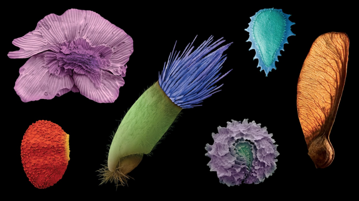
SEM scans of various plant seeds colorized
Supawat’s Looking Outwards Post
This week, I’d like to address Supwat’s Looking Outwards post from Week 5, where he writes about the magical microscopic universe uncovered through colored electron microscopy (SEM). It was really interesting to read an alternative perspective on one of the Miller Gallery’s most exciting exhibitions (in my opinion), World Within, created and curated by Rob Kesseler. What was most surprising to me when observing the artifacts and documentation of this photographic process is that SEM scans are taken only in black and white.
Here’s how it works: Through a an electric light filament, the machine shoots a flow of electron through a pair of electromagnetic lenses. The beam is varied and scans itself across the studied object. Depending on the position of the beam, an electron detector collects data from the secondary electrons that are repelled back from scan. After that, the image is run that through some filters that result in a extremely high resolution black and white image.
While these images are quite impressive on their own, without the sensibility of the artist who colors and shades these images, we wouldn’t be able to experience such evocative and beautiful colored imagery. It’s refreshing to find this kind of artistic collaboration even in the most dense and complex scientific studies.
Here is the tutorial Supawat linked that reveals the methodology behind coloring SEM scans:
Tutorial on Coloring SEM Scans
I took a gander and was truly blown away 🙂