Electron Evidence

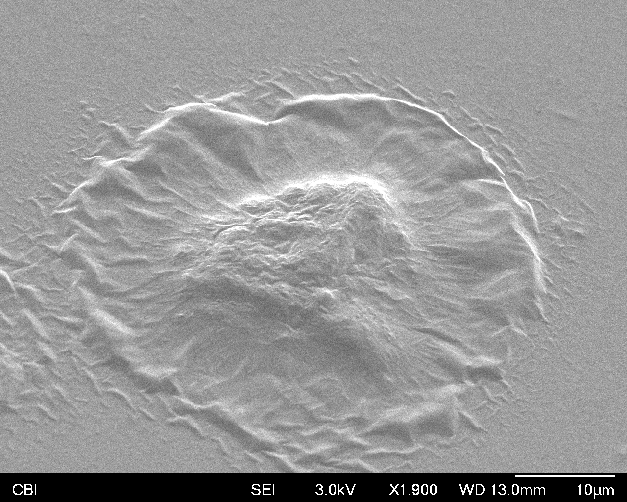
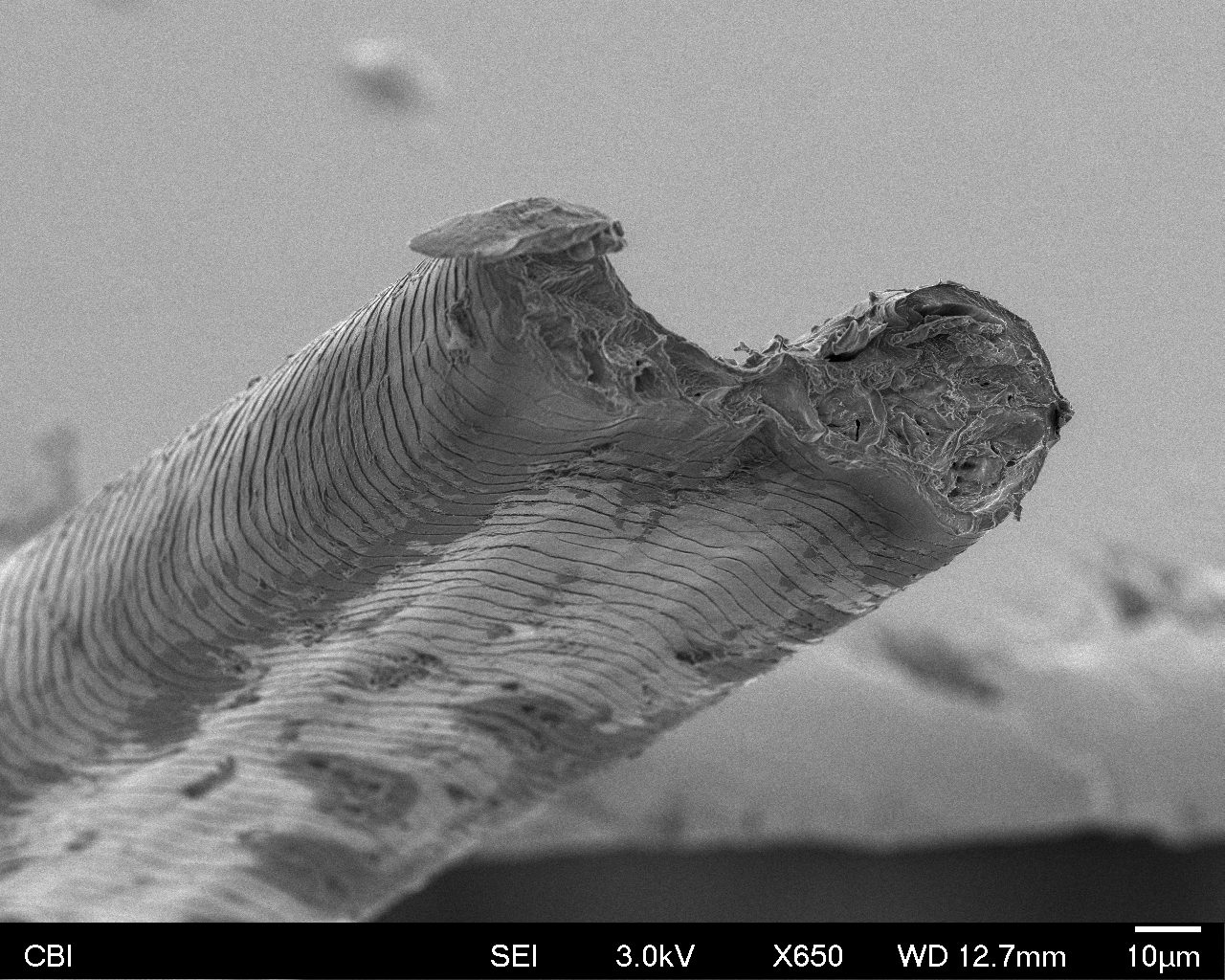
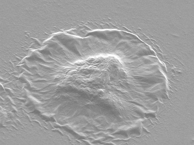
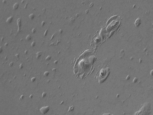
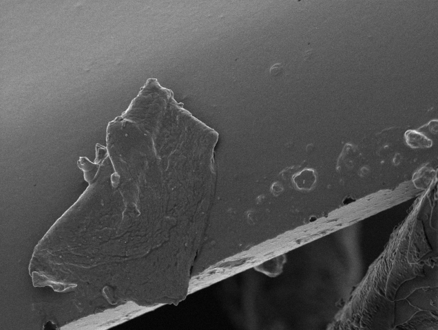
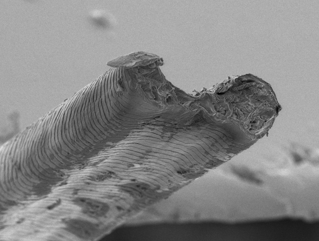
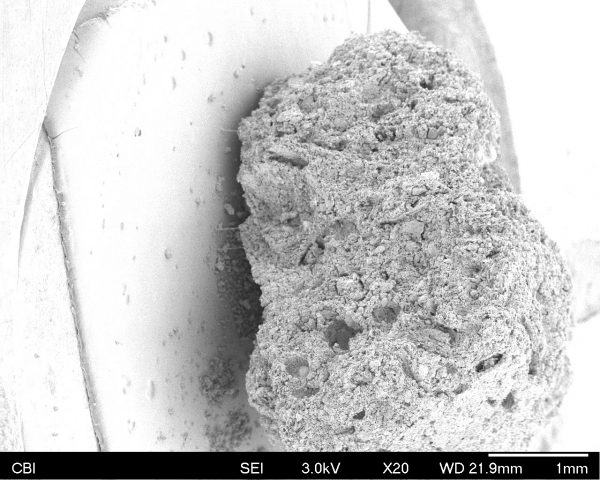
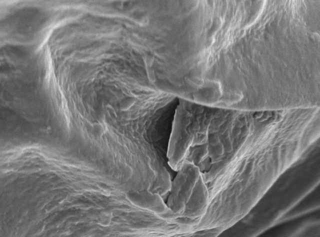
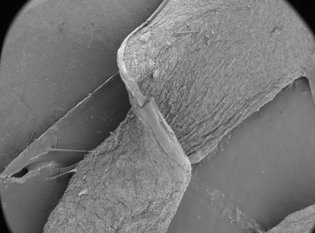
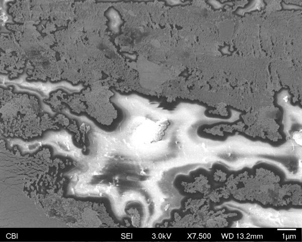
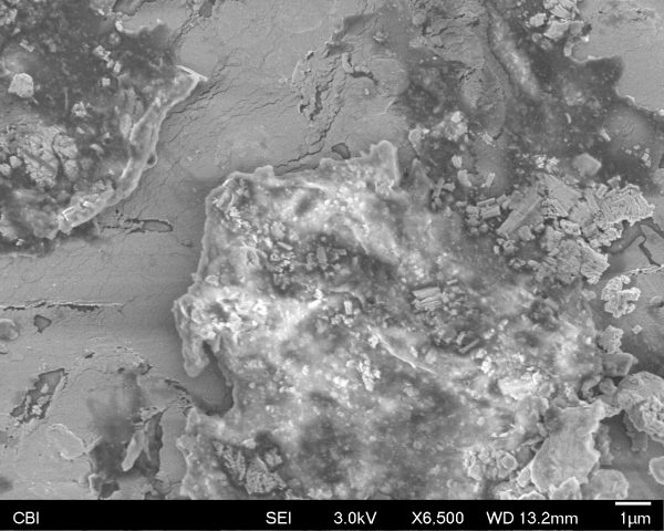
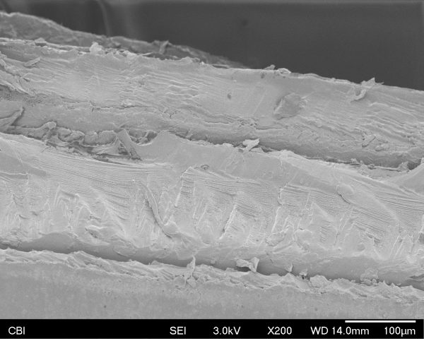
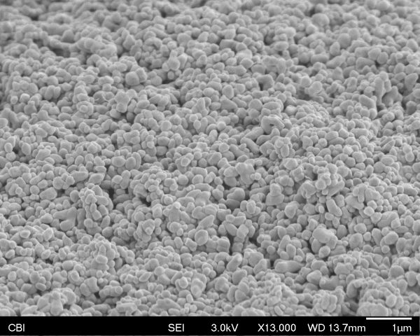
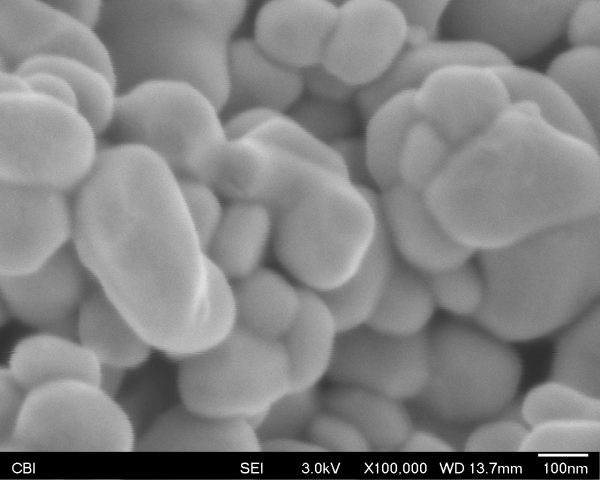
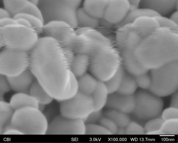
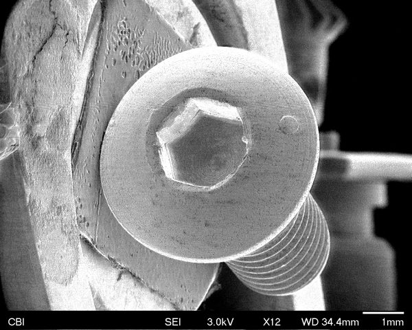
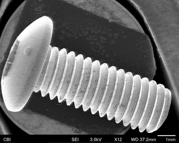
I scanned a screw that holds microphones and low resolution infrared cameras into my office’s wall. These devices were installed in my department’s offices, often without asking for consent of the occupants. I removed the whole sensor at one point, but got in a lot of trouble. Instead, this time I removed a screw and scanned that. The screw is unique, so this is “evidence” of my “crime”, in that it is evidence that this specific screw was removed from my office wall, and put into a fairly rare kind of microscope.

MarthasCatMug – SEM of a drain fly

The object I brought was a bug that I found out is actually a drain fly. They are super fuzzy and it was really cool to see how there were almost no surfaces, at any magnification, that did not have furry looking structures.

One place that was not fuzzy were the eyes, Donna explained to me that each individual ball is an eye and that the surface irregularities were actually an artifact of the fly being dried out.

Here is an image of the wing, due to the fly not being flat, a lot of the images ended up being of a very narrow depth of field.

You can see a bacteria on one of the fly’s feet! c:

Polaroid under SEM

The object I scanned was a Polaroid. I worried that the resulting images would be repetitive, but they turned out to be surprising.

The sputter coating on polaroid surface:


Edges of layers inside polaroid:


The silver grains coated on the film base:

At the magnitude of nanometers, the slight vibrations of the particles caused by sounds and shakes can be observed.

I was fascinated by how SEM captures the photos: it continuously captures a relatively long period. I like to think about how one single photo generated by SEM becomes a representation of a piece of music/ a conversation/ a series of actions.

Slight vibrations when a conversation was going on:

Vibrations when playing a piece of rock music:

Vibrations when knocking on the table:

kitetale – SEM

I brought a piece of ear plug that I used at a woodshop once. Since it was much bigger than what the machine could take, I ripped a portion of it. The result was pretty surprising — I was expecting a visual of foam, perhaps something like a sponge since that is how ear plugs contract and expand. Upon a closer look, it actually looked very flaky, as if it was made out of multiple thin layers of something.

I also had a chance to look at others’ objects too, which was quite a learning experience. This is a pollen found on one of the bugs:

SEM – Bee

All of my captures are of a bee. 

It was pretty interesting how this bee only had one type of pollen found on it, even though there are so many different types that exist. I also was able to see the bee’s second pair of eyes (on its forehead) which is something I never really noticed before. 

There were two photos I took of the same shot with a change in focus:

Found some pollen:

The bee’s hair looked like grass:

It was pretty surprising to go into this experience knowing close to nothing about bees and coming out of it knowing about their diet and anatomy through only magnified visual observation.