Today’s Agenda
- Selections from Shining360°
- Responses to the 1/14 Exit Ticket
- Continued Presentation: Some Inspirations
- Continued Presentation: Some ExCap Student Projects
- Main Lesson: Multispectral Imaging (part 1)
- Available multispectral equipment
- Hands-on: Multispectral portrait capture
- Discussion of Homework for 1/21
- Exit Ticket
Homework for Tuesday 1/21
Preparation for Claire Hentschker’s Photogrammetry Workshop (1/21):
- On your laptop, install MeshLab, an open-source viewer for 3D models
- On your laptop, install the Agisoft Metashape 30 day free trial
- Bring in an “object for a still life”
- Skim over this book about Still Life for inspiration
Preparation for Donna Stolz’s Scanning Electron Microscope (SEM) sessions (late January):
The Pitt Scanning Electron Microscope can display objects down to just a few nanometers in size, such as a virus. This is an utterly remarkable device. To prepare for your session, I ask you to please do the following, beforehand:
- On Tuesday 1/21, using the provided container, bring in a dry item, smaller than a pea, for the Scanning Electron Microscope. We recommend organic objects or materials. Donna will prepare your specimen.
- Before 1/21, sign up for a 25-minute session using this Doodle poll. Choose one option. Slots are first-come, first-served.
- Skim through the Wikipedia article about SEM imaging (3-5 minutes)
- On the website of the Pitt Center for Biologic Imaging, take the “Science 2019 Image Quiz” (5-10 minutes)
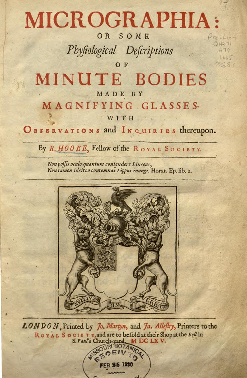
- Robert Hooke was the first person to make scientific observations under a microscope. Read four pages (which were originally numbered 112-116) in Robert Hooke’s 1665 classic, Micrographia, a historically significant book about his discoveries of the micro-world. In particular, You are asked to read the 4-page section, Observation XVIII, “Of the Schematism or Texture of Cork, and of the Cells and Pores of some other such frothy Bodies”, in which he describes, for the first time ever in scientific literature, the discovery of cells. (This is the passage in which the biological term “cell” was coined.) Don’t be thrown by Hooke’s use of the “long s” ( ſ ), which is an archaic form of the lower case letter s that fell out of use around 1800. Below are links to three different versions of Micrographia. Please note that Hooke’s original pagination appears differently in each of the following PDFs. The pages you should read, which Hooke’s printer numbered 112-116, appear as:
- PDF pages 155-160 in this best-quality original scan
- PDF pages 174-180 in this easily downloadable PDF scan
- PDF pages 131-135 in this easiest-to-read OCR plain text