For my visit to the Pitt Center for Biologic Imaging I cut a tiny slice of a dried sweetened orange slice.
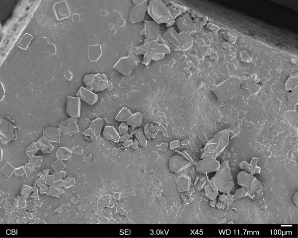
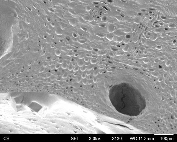
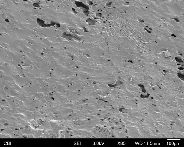
millimeter scale (above)
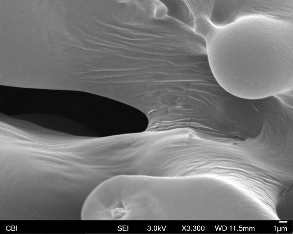
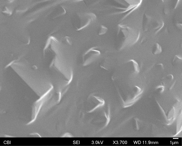
nanometer scale (above)
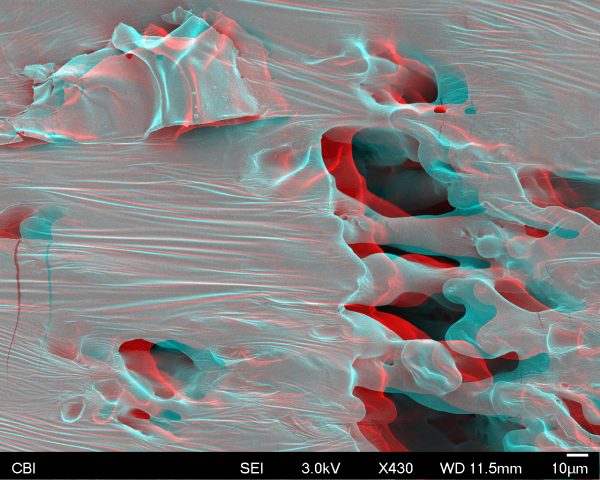
anaglyph (above)
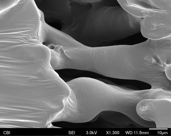
I was really blown away by this process! It was really fascinating to be able to see something already so tiny, even further up close. The larger squares in the first image are sugar crystals. In the more zoomed in images, it appears to look like a weird, swiss cheese moonscape. Upon my arrival I learned that there was some concern that this item would not work, as there may have been small amount of moisture in even the dried fruit, but it did (Donna tested this and knew this before my arrival). I am looking forward to seeing what other people captured. In my visit we discussed what objects/processes and how photogrammetry might work with these types of images. We ended up discussing what bugs might look like under the Scanning Electron Microscope and then turned into photograms.
Other photos below: