SEM-Tagbamuc

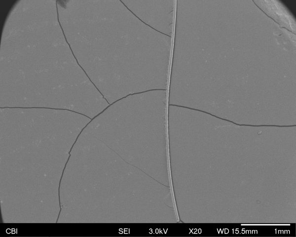
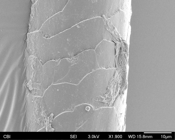
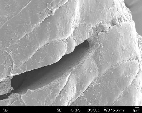
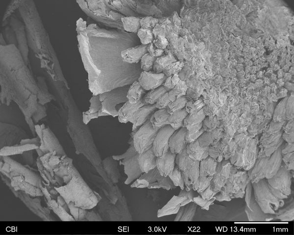
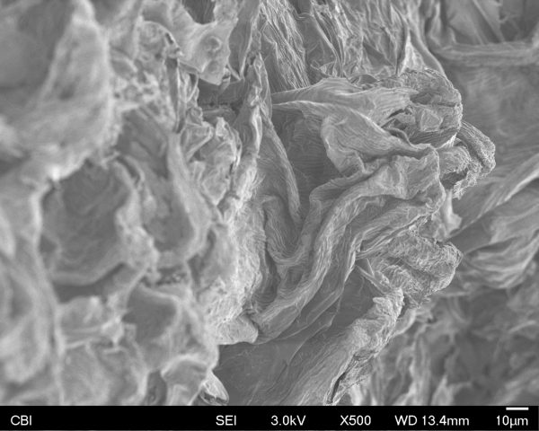
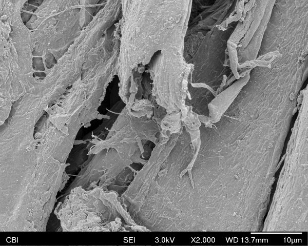
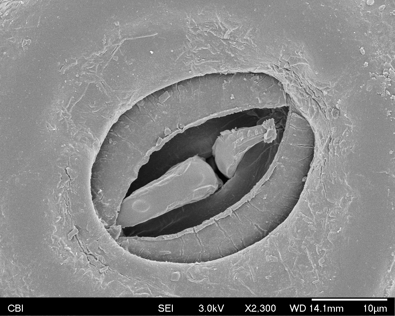
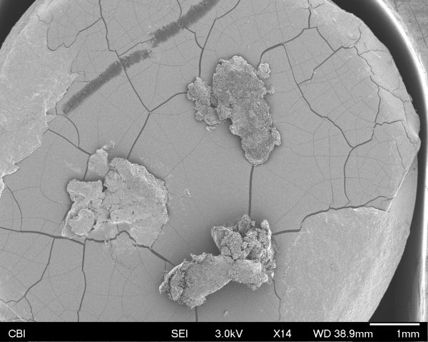
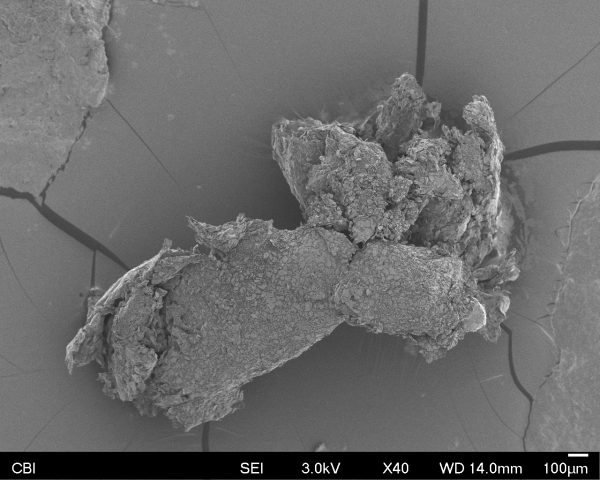
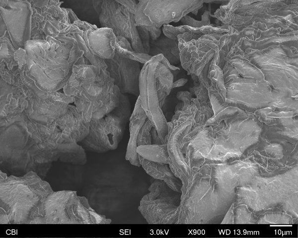
Please find my SEM results bellow of a piece of dry skin. For reference, these flakes are small enough to fit on a head of a pin;
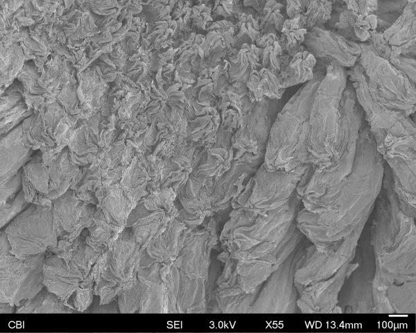
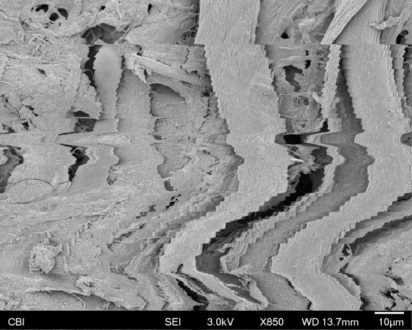
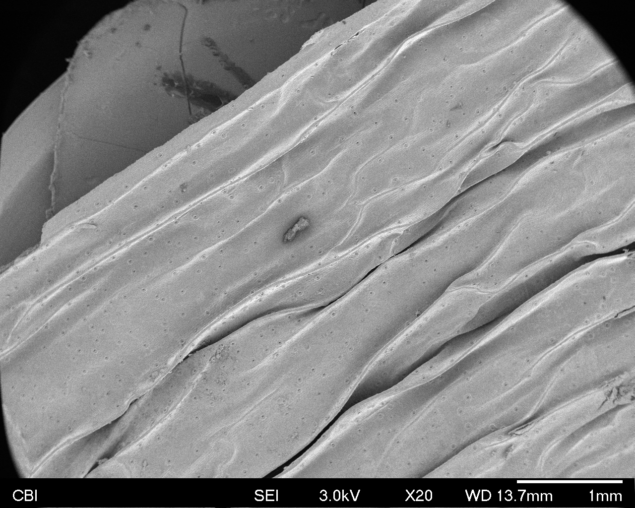
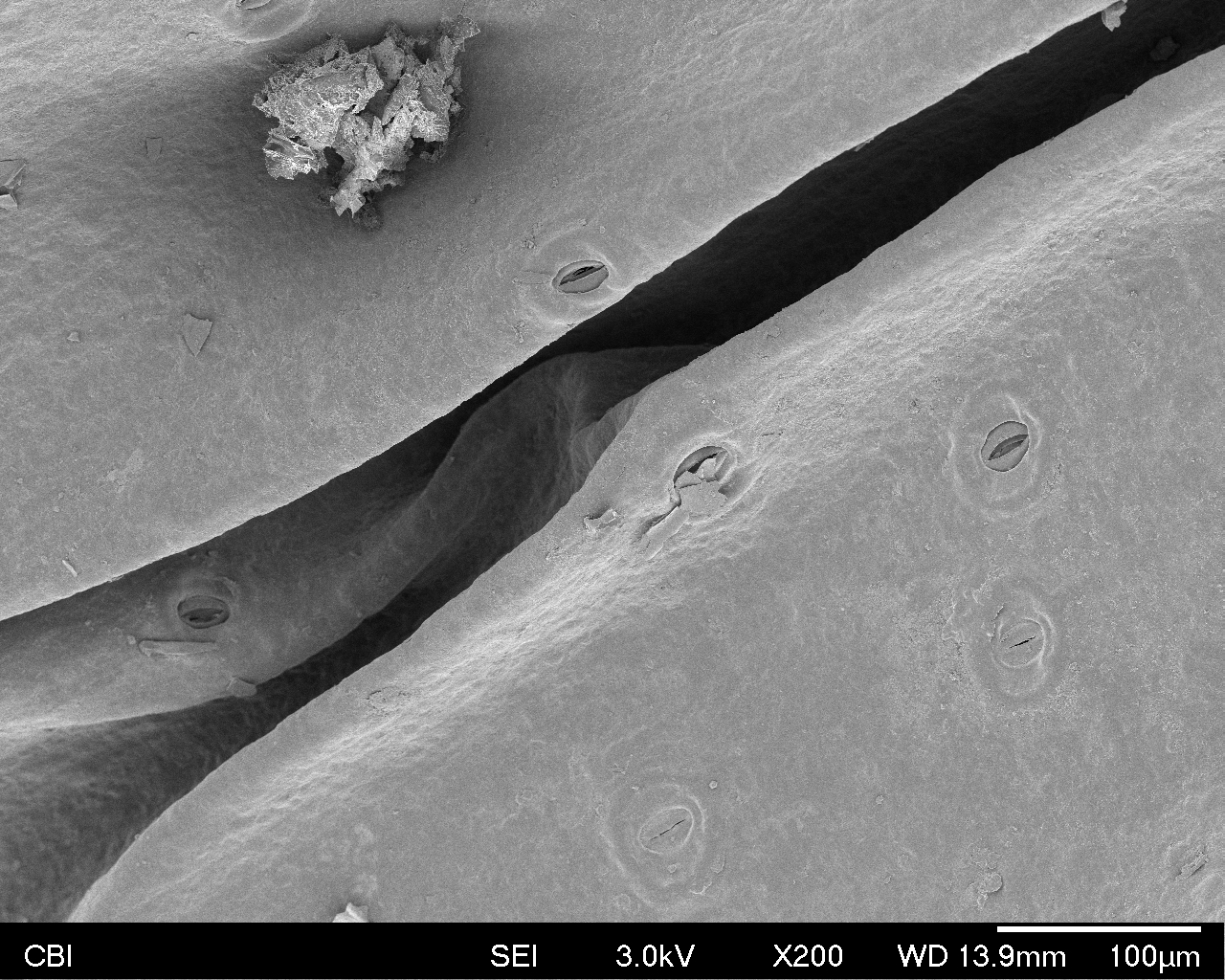
Familiar view;

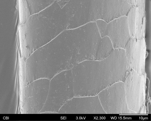
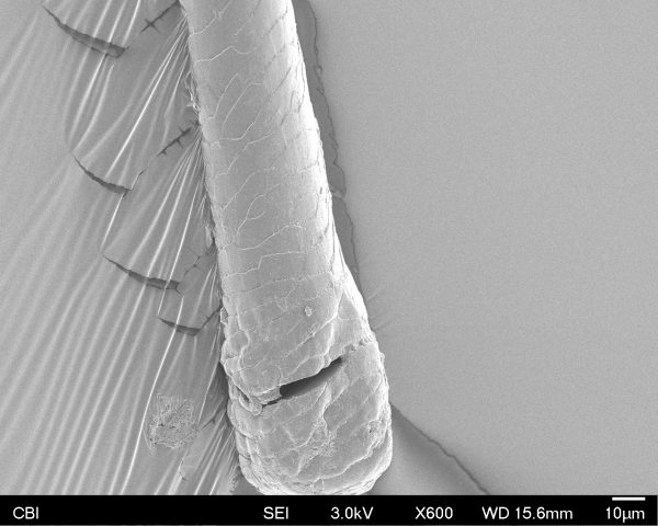
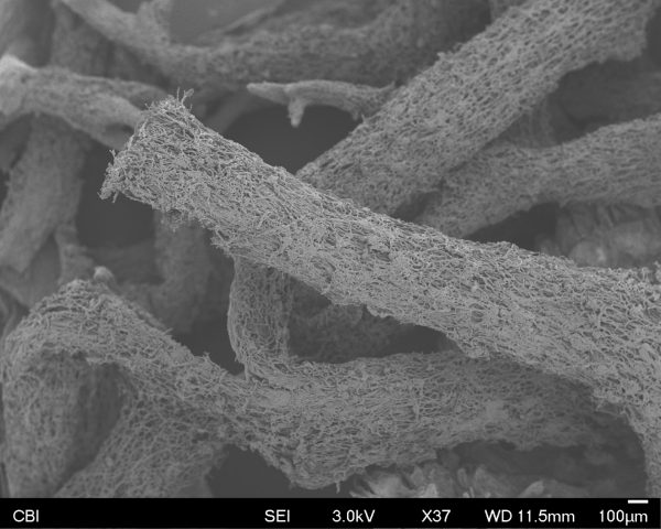
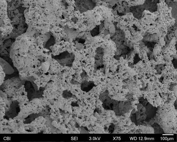
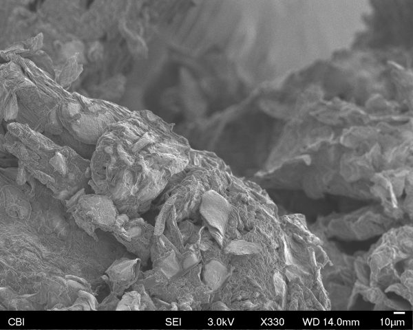
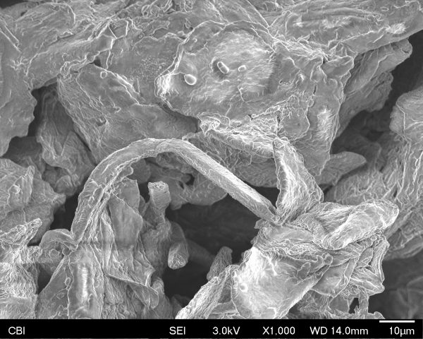
Detail shots/Unfamiliar Views
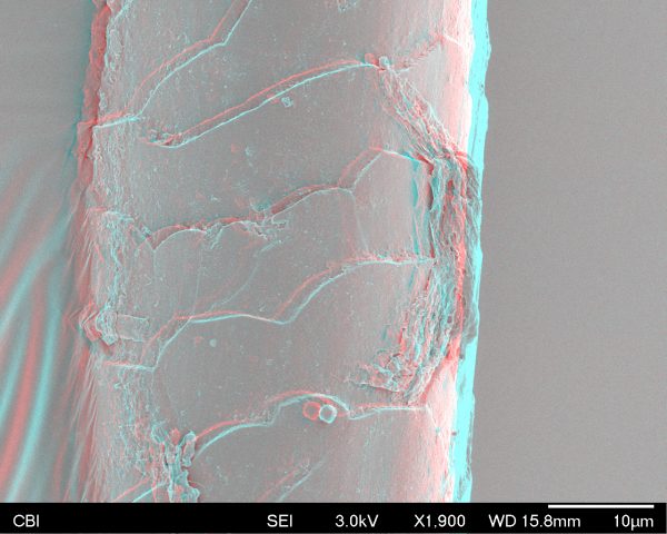
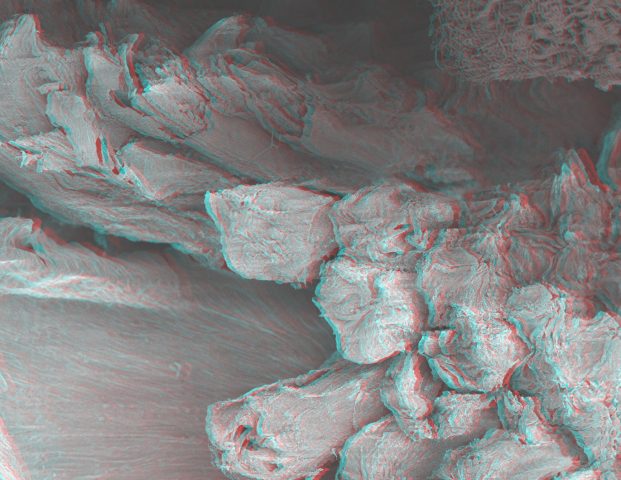
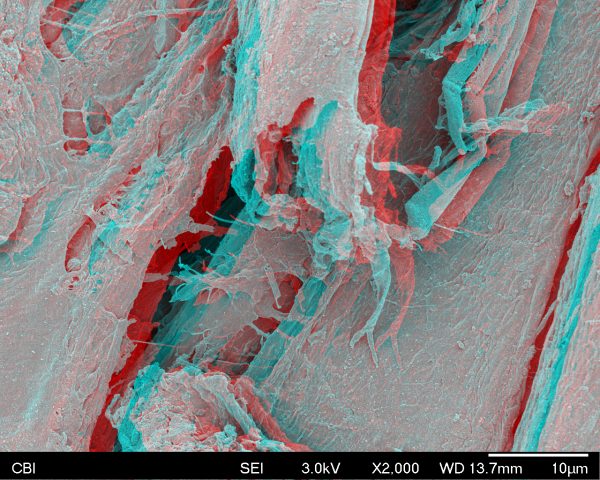
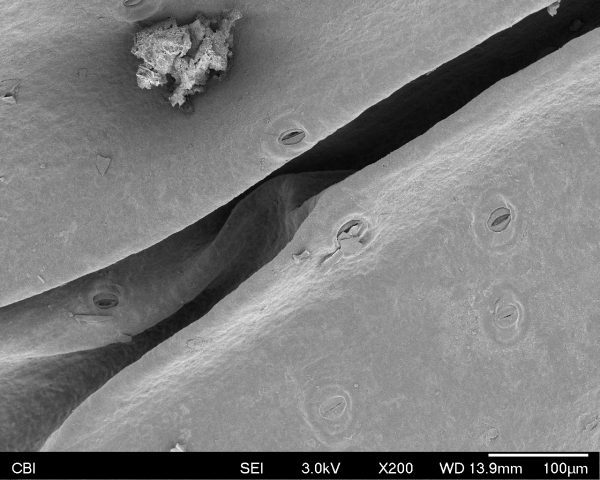
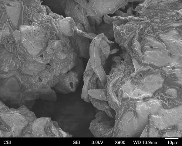
StereoPairs

Experience 

Donna can testify that I was really surprised and delighted by my experience. I think what caught me so off guard was how layered dead skin is and how it interacts with each other. The first thing I thought when I was looking at it under the microscope, is that it looks like a tiny cave where some kind of creature should live! The flakes definitely inspired me to design a world and a creature to exist in this space. It was pretty mind-blowing to think that came off of my body and has so much detail for something that was just a spec on my clothes.

Donna was telling Nica and I that it would be difficult to get the stereopairs done correctly because of where my sample was located on the disk. Apparently some positions favor the process more than others, but we gave it a shot anyway!

Workshop – SEM

The Scanning Electron Microscope was an interesting experience to observe the minuscule world around us. I brought in an unknown white hair that I found on the floor of the Chroma Key Studio. I thought it was synthetic or from a coat, but I discovered that it was most likely a human hair due to its secretion structure. It was also determined it was not a “black” hair due to its composition (so it was not mine) and that it probably fell since it still had an intact bulbous base. I found it intriguing that this could also be decided from close evaluation of its physical form. Along with the required images, I also captured a spot with hair product potentially on it and a break in the middle of the hair.

Familiar View (millimeter scale)

Unfamiliar View (nanometer scale)

Stereo Pair


Additional Images:


SEM

In the vial I combined a fragment of moss and chamomille – not the petals of the flower but the stigma, the head of the pistil.

The first image shows a familiar view from the chamomile filaments on the left side and moss on the right side of the image. I was mesmerized by the detail of the filaments. In some areas the moss looked like tree bark in others it was very porous, like a sea sponge.

The second and third image is a close up of the chamomile filaments and the fourth one is of the fragment moss where you can really see its sponge-like features. It was the first time I used a scanning electron microscope and though I expected to see this type of visuals, I could not help feeling marvelled with every image.

What interests me visually is the softness and tenderness of the images. I am drawn into its graphic quality, they seem very delicate graphite drawings.

SEM – burnt matchstick

My object was a burnt matchstick. The first image was the tip of the matchstick but unfortunately it broke off in the microscope which meant it was no longer grounded and was harder to focus at higher mags. The Second image was a result of the more receptive surface of the matchstick for the microscope. Having Donna guide me through the process of how the microscope worked was fascinating and very exciting to see that the image is a raster. This ultimately led me to do the fun trick you can do on a photocopier. By moving the source or the camera as the image begins to populate you can stretch and warp the image. By moving both the joystick and the angle of the sample plate I was able to come out with a very different image (Image 3). Image 4 is the anaglyph pair.


SEM Results

I scanned a piece of a dried leaf that I picked up from my dying houseplant. At x20 magnitude, we are able to see veiny look of the leaf as well as small openings on the surface. I learned from Donna that these openings are called ‘stomata.’ A stoma is like a pore that facilitates gas exchange for plants. At x2,300 mag, we are able to see it more clearly — a few different cells act as “doors” to control the open and close of the “pore.”

Donna said that the SEM that we used is around fifteen years old. The combo of the analog and digital interfaces made it seem like a highly functioning piece of historical artifact. Navigating it also felt like remotely controlling a robot in another planet.

Due to my complete lack of knowledge in biology, I definitely did not expect to see the stoma and the even smaller cells. I did expect magical, unfamiliar patterns, but I didn’t know that this act of ‘seeing’ would lead to a more visceral understanding of the underlying structures, actions, and ‘stories’ of life. Now I can better understand what Robert Hooke might have felt when he saw those cells under his hand-crafted microscope.

Below: stereo pair