Shrimp tail SEM scanned pics

The object I scanned is a shrimp tail(cooked cuz I took it from my thai fried rice). The first pic is the feathers that shrimp used for sensing water flows but with grease on it. The second pic i found it so amazing is that it is the broken joints of the feathers on the tail and it even looks like animal skeleton(or teeth).

shrugbread-SEM

For this trip to CBI’s Scanning Electron Microscope I brought in a couple flakes of fish food hoping to see tiny bits of organic matter however preserved in the flat flake. This project is how I actually learned that fish food is only part finely powdered”fish-meal” with the rest of the concotion just being a soup of wheat, letchin, and water.

I opened with this and it was promising to have really interesting caves and craters to explore

But due to how thin and shallow the craters are they stop showing a lot of clarity. This was still incredibly insightful! The most fun part of this is using the controller that sets your framing and zooming, it’s a very different frame of mind than digital photography.

hunan – SEM

The two objects I chose are a light roast Ethiopian coffee bean and a piece of developed BW 400 film. The film is pretty boring — just a flat surface. According to Donna, the silver particles are apparently too small for the SEM. But the coffee was super interesting with a variety of textures and internal structures. The images below are focus stacked from 8~15 shots.

SEM Experience


It goes without saying this experience was incredible… but I’ll say it anyways. We were told about the amazing resolving power of the tiny image… but I was surprised by how we were able to focus at every resolution.  I was also surprised by my own desire to keep zooming out to try and see the whole object/orient myself in the image.

This is a .gif of the tip of a discarded cat’s claw ft. a little bit of dirt on the tip.

This is a picture of the joint between a fly’s wing and it’s body.

bumble_b-SEM

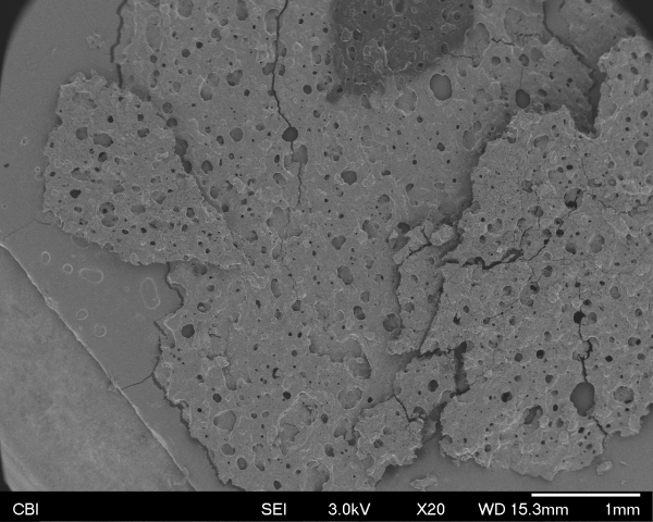
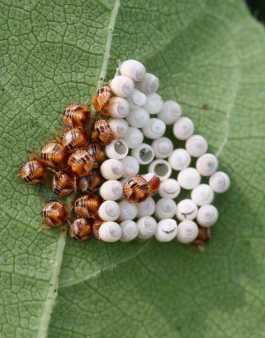
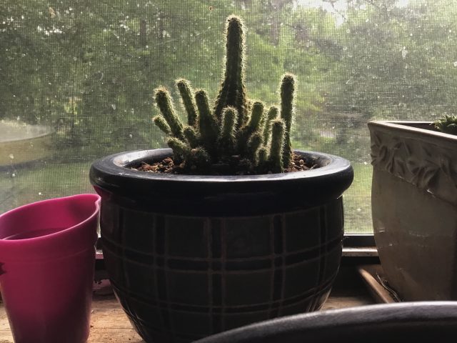
I brought in two samples, one of my beloved cactus Peanut, and one of a bug I found dead on my wall that broke into pieces the moment I picked it up.

Here is a photo of Peanut from over 2 years ago when I first got him. (Home Depot treated him poorly, so he looks terrible in this, but he is much healthier now.)

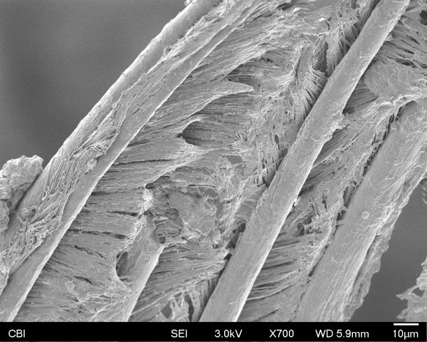
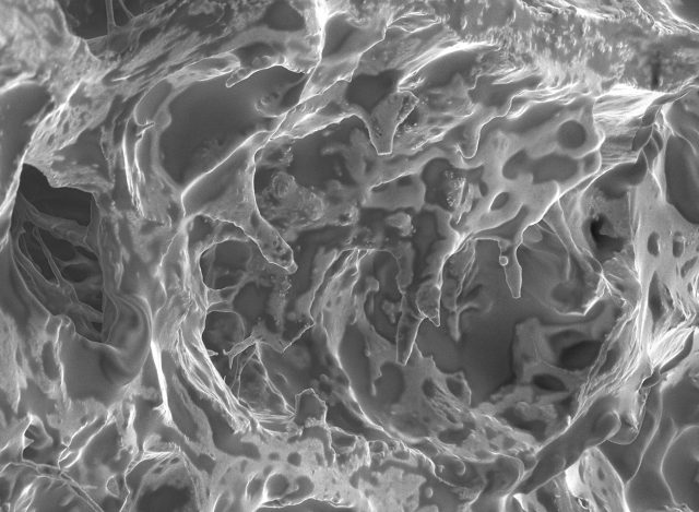
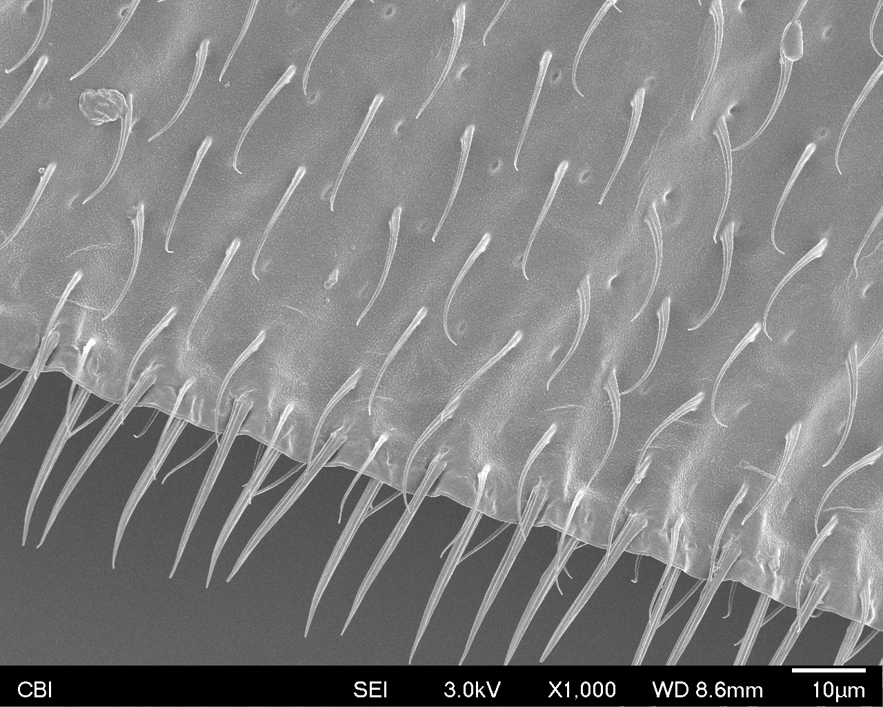
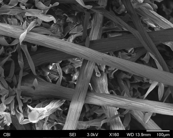
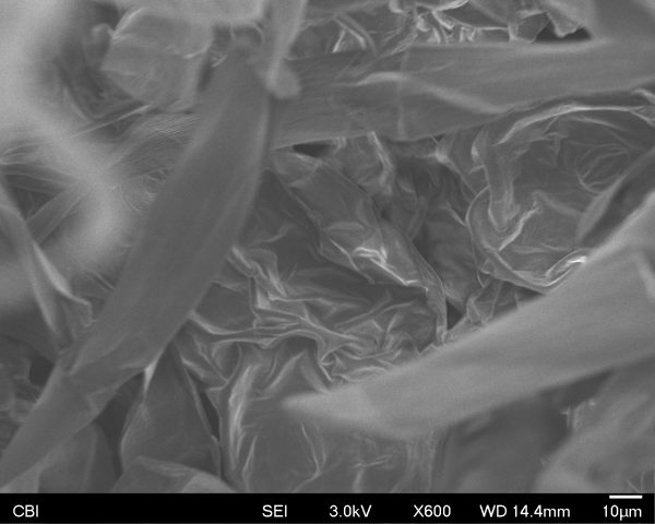
Here is how a small tip that I chopped off him looks under the microscope:

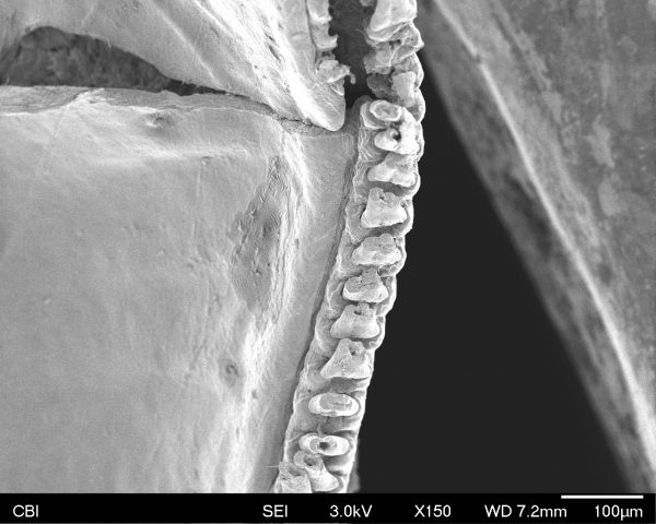
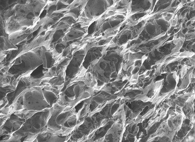
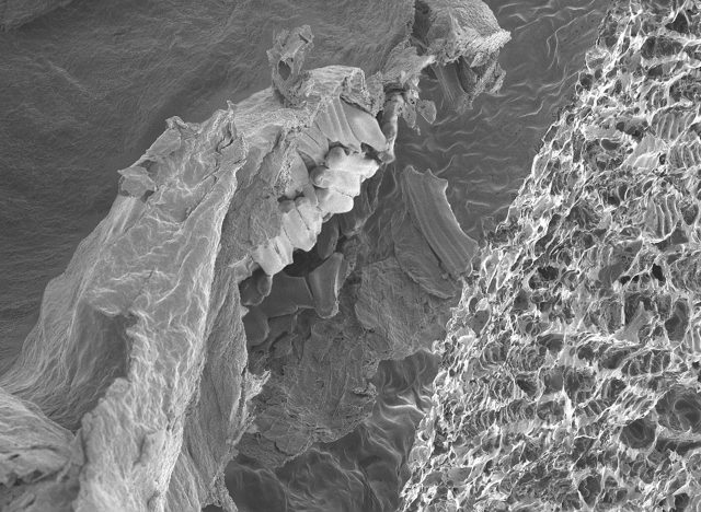
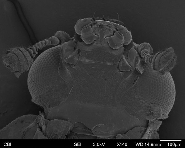
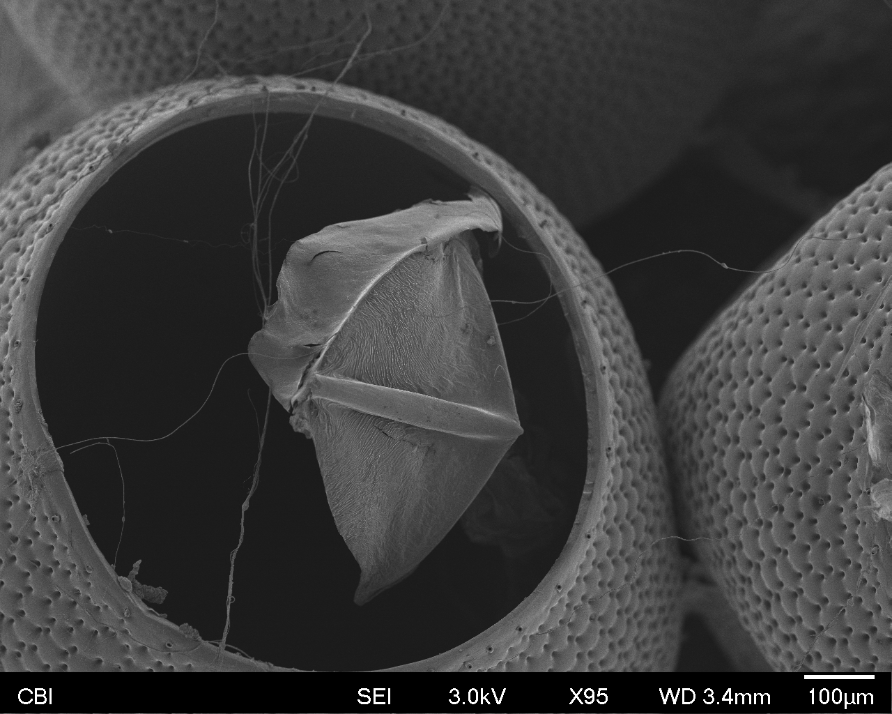
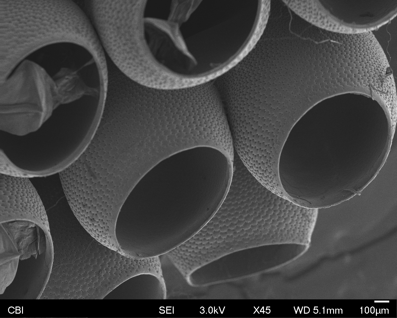
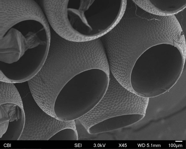
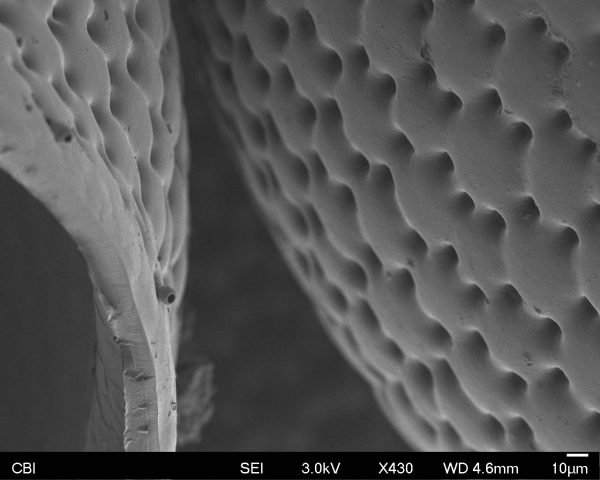
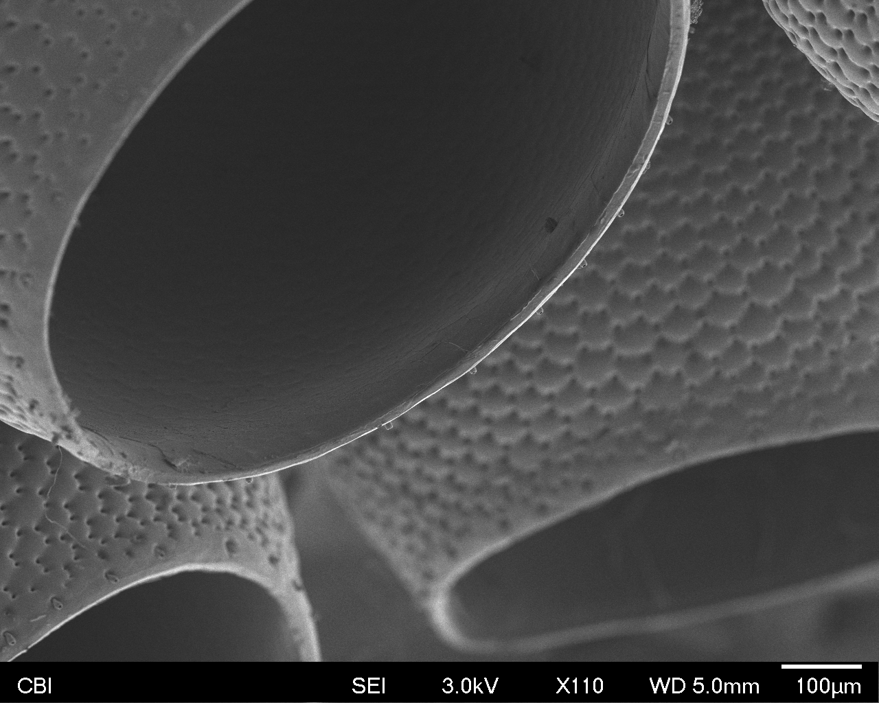
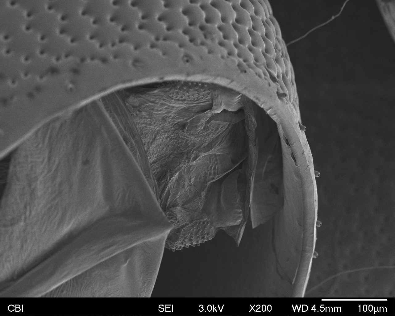
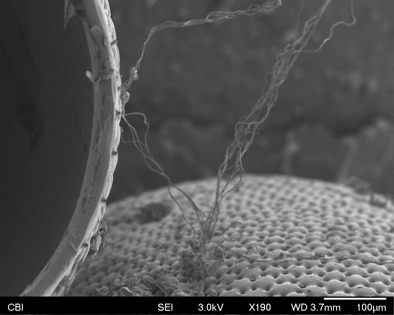
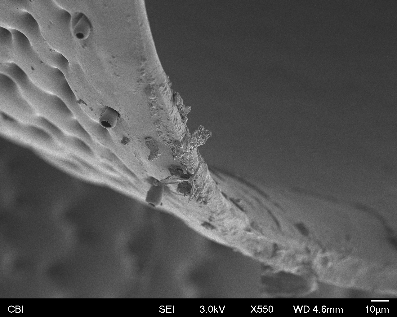
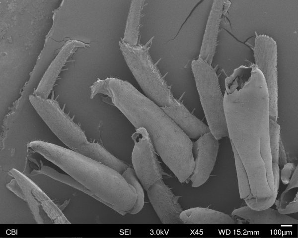
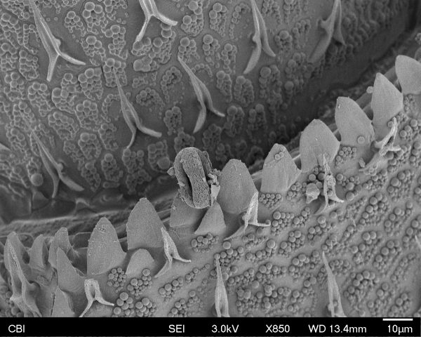
My favorite thing about the bug I found is how there are so many holes in his body from where all his limbs fell apart. It’s actually kind of sad, but I’m happy to know it at least happened after he died. You can see those holes here:

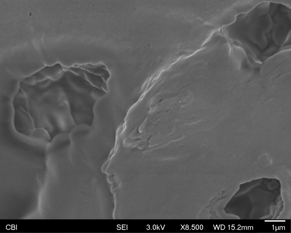
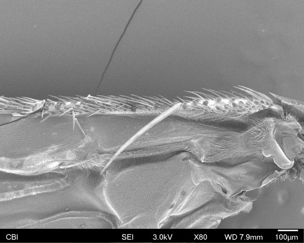
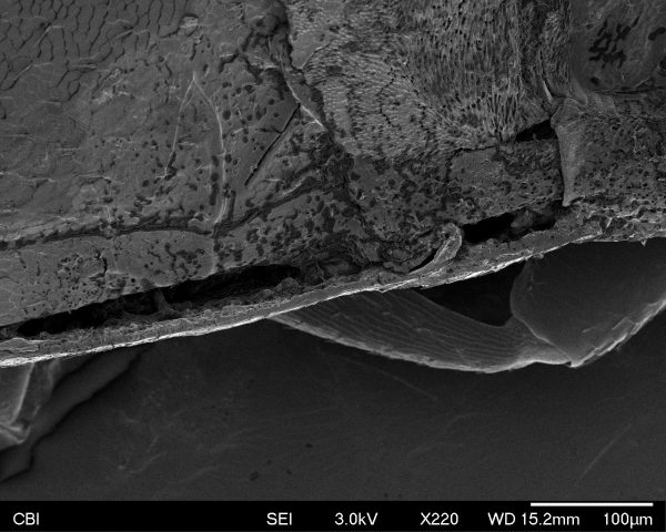
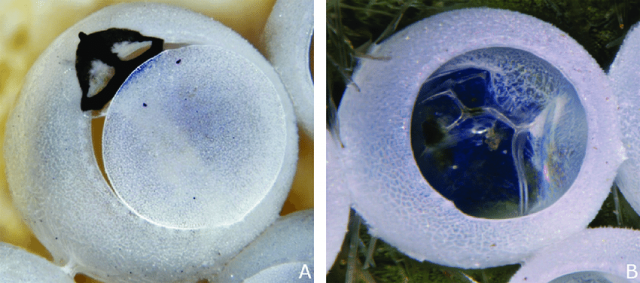
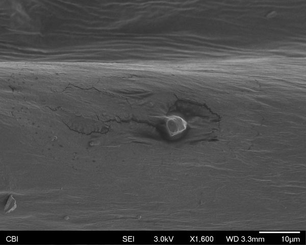
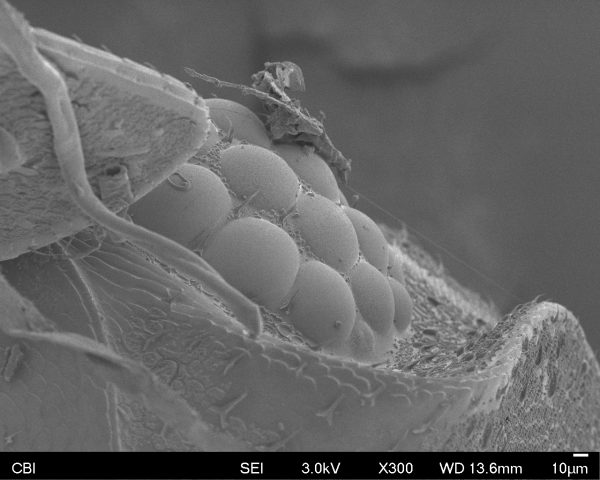
Here is his eye (with a little piece of guck):

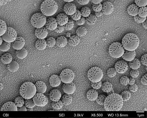
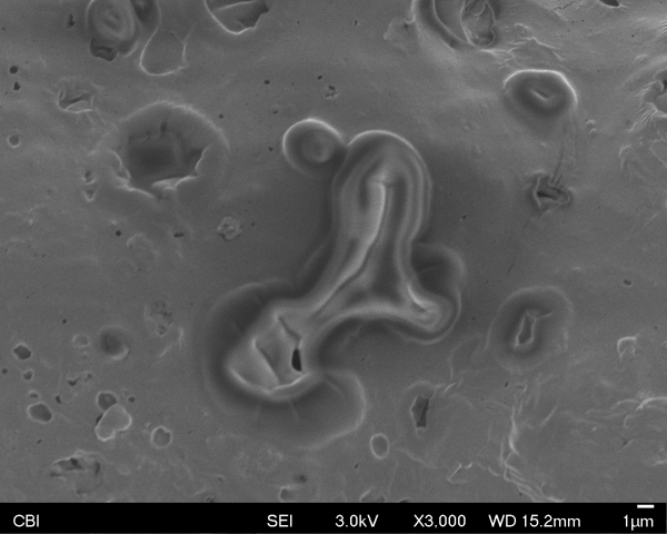
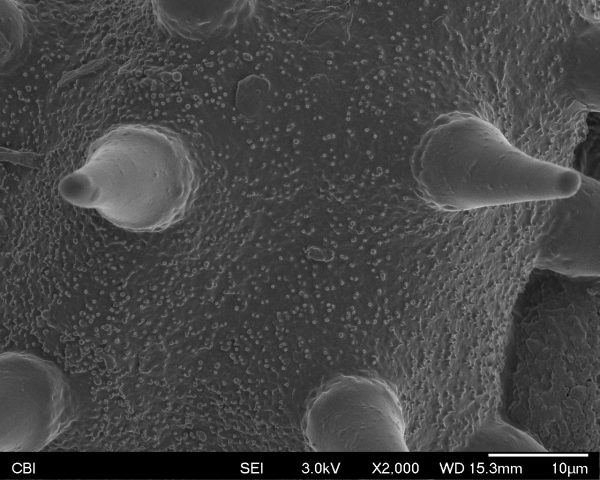
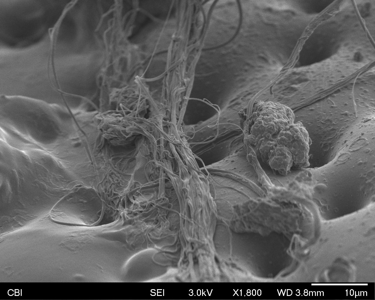
Here is a piece of pollen we discovered on his body:

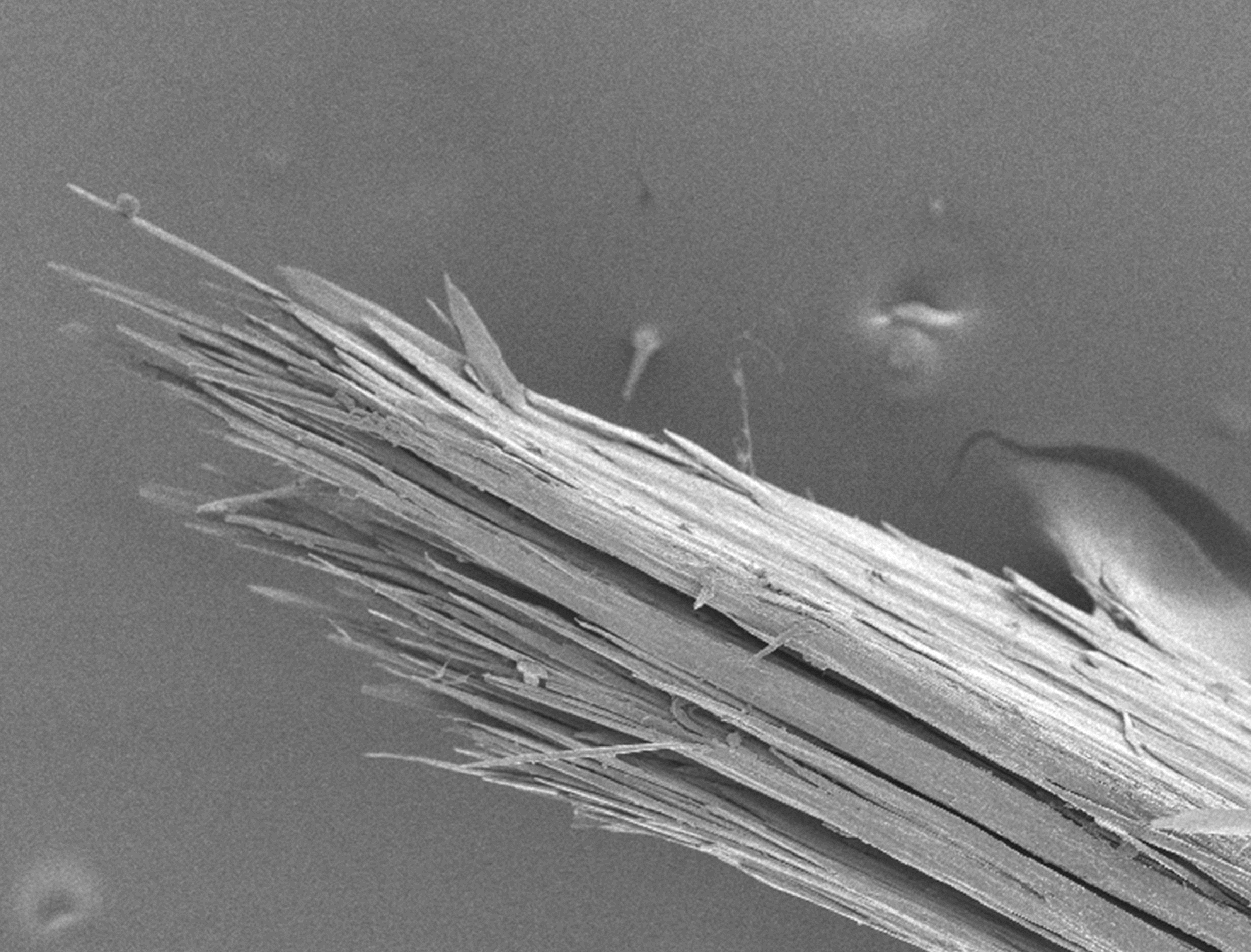
And here is Donna’s favorite photo that we found at the way end. She was so excited that she made herself a copy:

marimonda – SEM

In the electron microscope, I scanned three different types of bugs and pollen. I literally put the taped magnets inside of a flower to collect different organisms and collected them in the tube without much concern. So my first surprise was actually finding out the number of bugs and pollen I ended up collecting. The second thing that interested me, was the complexity of the organisms, and the places where you zoomed so far in that you didn’t even recognize the original subject (where your brain filled in landscapes/context for the unrecognizable images).